产品描述Phosphatidylinositol 3-kinase (PI 3-kinase) is composed of p85 and p110 subunits. p85 lacks PI 3-kinase activity and acts as an adapter, coupling p110 to activated protein tyrosine kinase. Two forms of p85 have been described (p85α and p85β), each possessing one SH3 and two SH2 domains. Various p110 isoforms have been identified. p110α and p110β interact with p85α, and p110α has also been shown to interact with p85β in vitro. p110 expression is restricted to white blood cells. It has been shown to bind p85α and β, but it apparently does not phosphorylate these subunits. p110δ seems to have the capacity to autophosphorylate. p110γ does not interact with the p85 subunits. It has been shown to be activated by α and β γ heterotrimeric G proteins.
WB: 1:1,000
IHC-P: 1:50-1:100
FC: 1:50-1:100
IP: Use at an assay dependent concentration.
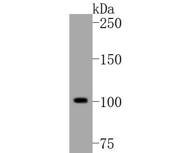
Fig1: Western blot analysis of PI3-kinase p110 subunit beta on Hela cell lysates. Proteins were transferred to a PVDF membrane and blocked with 5% BSA in PBS for 1 hour at room temperature. The primary antibody (ET1610-36, 1/500) was used in 5% BSA at room temperature for 2 hours. Goat Anti-Rabbit IgG - HRP Secondary Antibody (HA1001) at 1:5,000 dilution was used for 1 hour at room temperature.
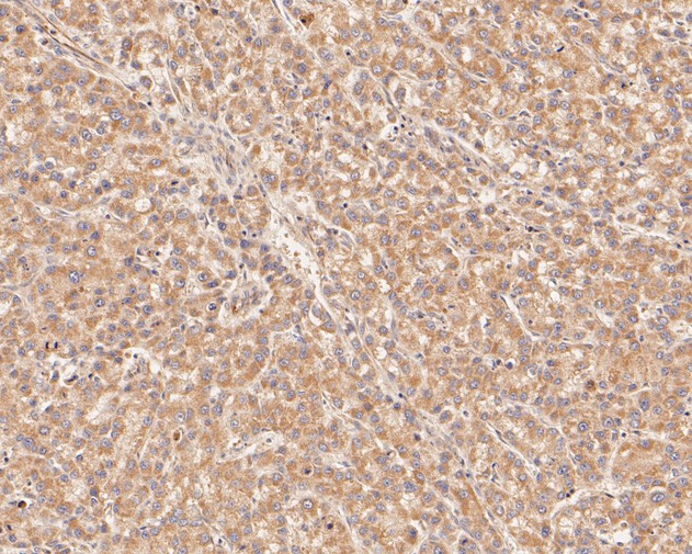
Fig2: Immunohistochemical analysis of paraffin-embedded human liver carcinoma tissue using anti-PI3-kinase p110 subunit beta antibody. The section was pre-treated using heat mediated antigen retrieval with Tris-EDTA buffer (pH 8.0-8.4) for 20 minutes.The tissues were blocked in 5% BSA for 30 minutes at room temperature, washed with ddH2O and PBS, and then probed with the primary antibody (ET1610-36, 1/50) for 30 minutes at room temperature. The detection was performed using an HRP conjugated compact polymer system. DAB was used as the chromogen. Tissues were counterstained with hematoxylin and mounted with DPX.
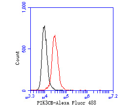
Fig3: Flow cytometric analysis of PI3-kinase p110 subunit beta was done on Hela cells. The cells were fixed, permeabilized and stained with the primary antibody (ET1610-36, 1/50) (red). After incubation of the primary antibody at room temperature for an hour, the cells were stained with a Alexa Fluor 488-conjugated Goat anti-Rabbit IgG Secondary antibody at 1/1000 dilution for 30 minutes.Unlabelled sample was used as a control (cells without incubation with primary antibody; black).
背景文献
1. H land K et al. Targeting class IA PI3K isoforms selectively impairs cell growth, survival, and migration in glioblastoma. PLoS One 9:e94132 (2014).
2. Wojtalla A et al. Targeting the phosphoinositide 3-kinase p110-a isoform impairs cell proliferation, survival, and tumor growth in small cell lung cancer. Clin Cancer Res 19:96-105


 我的购物车(0)


 成功收藏产品


 正品保障
正品保障 8000万+
 期货精准
期货精准 便捷价优
便捷价优 京公网安备 11010802021763号
增值电信业务经营许可证:京B2-20192263
ICP备案号:京ICP备14048343号-2
Copyright© 供应室版权所有
2007-2025,All Rights Reserved
京公网安备 11010802021763号
增值电信业务经营许可证:京B2-20192263
ICP备案号:京ICP备14048343号-2
Copyright© 供应室版权所有
2007-2025,All Rights Reserved

